Home

send taste Homeless light bulb sign mri Odds lonely accent

Appearance of Meningiomas on Diffusion-weighted Images: Correlating  Diffusion Constants with Histopathologic Findings | American Journal of  Neuroradiology
Appearance of Meningiomas on Diffusion-weighted Images: Correlating Diffusion Constants with Histopathologic Findings | American Journal of Neuroradiology

Presentation1.pptx, radiological classical signs and appearances in  neuroradiology.
Presentation1.pptx, radiological classical signs and appearances in neuroradiology.

Figure, Light bulb sign in cerebellar abscess in DWI MR image. Contributed  by Sunil Munakomi, MD] - StatPearls - NCBI Bookshelf
Figure, Light bulb sign in cerebellar abscess in DWI MR image. Contributed by Sunil Munakomi, MD] - StatPearls - NCBI Bookshelf

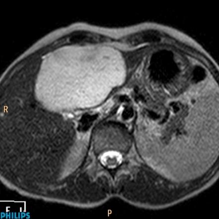
liver hemangioma MRI

Mark Mamlouk on Twitter: "@The_ASPNR @TheASNR @ASHNRSociety #Radiology can  play a big role in diagnosing these benign tumors if there is no cutaneous  component to a deep hemangioma. US is usually all
Mark Mamlouk on Twitter: "@The_ASPNR @TheASNR @ASHNRSociety #Radiology can play a big role in diagnosing these benign tumors if there is no cutaneous component to a deep hemangioma. US is usually all

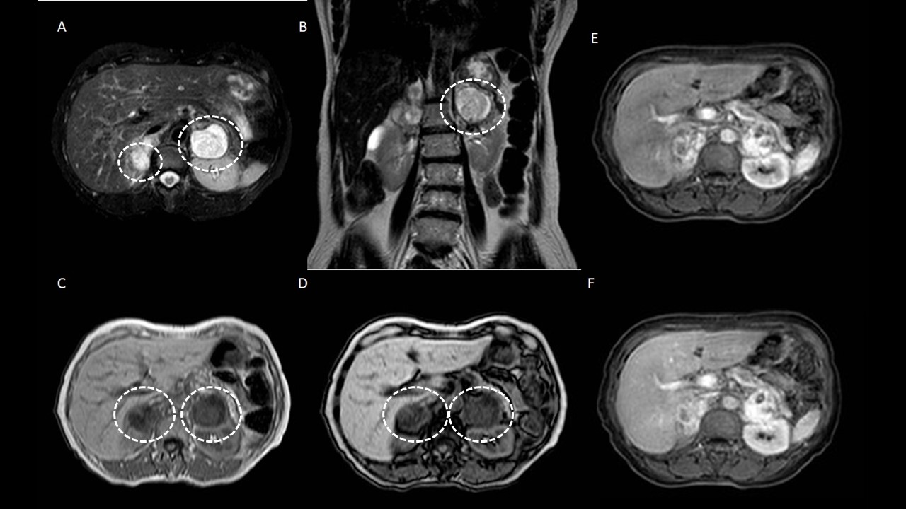
Pheochromocytoma: The mass is hypointense (arrow) on (a) axial... |  Download Scientific Diagram
Pheochromocytoma: The mass is hypointense (arrow) on (a) axial... | Download Scientific Diagram

Abdomen and retroperitoneum | 1.10 Adrenal glands : Case 1.10.3  Pheochromocytomas | Ultrasound Cases
Abdomen and retroperitoneum | 1.10 Adrenal glands : Case 1.10.3 Pheochromocytomas | Ultrasound Cases

Daniel J. Kowal, MD | Radiologist Headquarters on Twitter: "Multilocular  right endometrioma on MRI adherent to uterus (U). Homogenously light-bulb  bright on T1 (blue) & dark on T2 = “T2 shading” (orange),
Daniel J. Kowal, MD | Radiologist Headquarters on Twitter: "Multilocular right endometrioma on MRI adherent to uterus (U). Homogenously light-bulb bright on T1 (blue) & dark on T2 = “T2 shading” (orange),
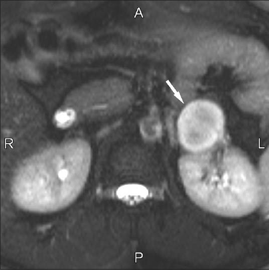
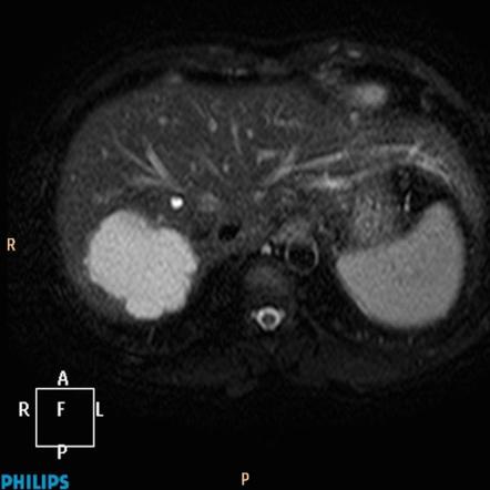
Hepatic hemangioma - with light bulb sign | Image | Radiopaedia.org

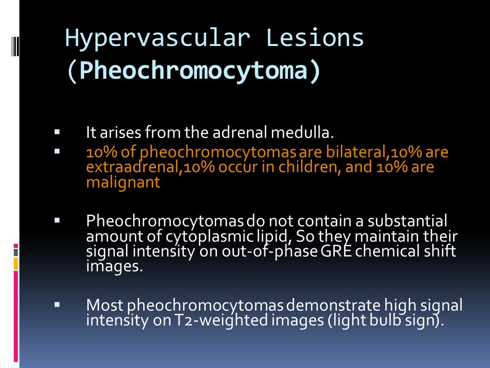
The light bulb sign in pheochromocytoma | SpringerLink

MRI of Adrenal and Extraadrenal Pheochromocytoma | AJR

The “light bulb sign” in liver hemangioma | SpringerLink

Liver haemangioma - with light bulb sign | Radiology Case | Radiopaedia.org

Adrenal Lesions: Spectrum of Imaging Findings with Emphasis on  Multi-Detector Computed Tomography and Magnetic Resonance Imaging - Journal  of Clinical Imaging Science
Adrenal Lesions: Spectrum of Imaging Findings with Emphasis on Multi-Detector Computed Tomography and Magnetic Resonance Imaging - Journal of Clinical Imaging Science

Adrenal Masses: MR Imaging Features with Pathologic Correlation - ppt  download
Adrenal Masses: MR Imaging Features with Pathologic Correlation - ppt download

MRI of Adrenal and Extraadrenal Pheochromocytoma | AJR

LIGHT BULB SIGN - Radiology Classroom | Facebook

Appearance of Meningiomas on Diffusion-weighted Images: Correlating  Diffusion Constants with Histopathologic Findings | American Journal of  Neuroradiology
Appearance of Meningiomas on Diffusion-weighted Images: Correlating Diffusion Constants with Histopathologic Findings | American Journal of Neuroradiology

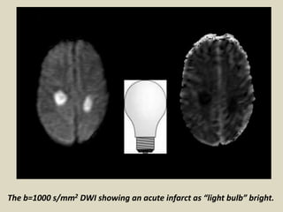
Pheochromocytoma MRI - wikidoc

PHEOCHROMOCYTOMA- MRI - YouTube

EPOS™
EPOS™

LIGHT BULB SIGN - Radiology Classroom | Facebook

Light bulb sign (hepatic hemangioma) | Radiology Reference Article |  Radiopaedia.org
Light bulb sign (hepatic hemangioma) | Radiology Reference Article | Radiopaedia.org

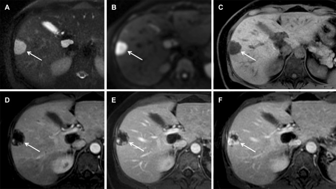
MRI of a typical hemangioma in liver segment VIII. T2-weighted imaging... |  Download Scientific Diagram
MRI of a typical hemangioma in liver segment VIII. T2-weighted imaging... | Download Scientific Diagram

Cavernous hemangioma A, T2-welghted MR image shows typical... | Download  Scientific Diagram
Cavernous hemangioma A, T2-welghted MR image shows typical... | Download Scientific Diagram

Figure 1 from Brain attack: a new approach to stroke. | Semantic Scholar